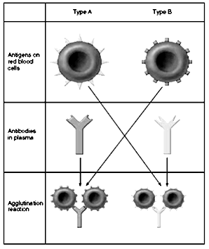
When blood is mismatched so that anti-A or anti-B plasma agglutinins are mixed with red blood cells that contain A or B agglutinogens, respectively, the red cells agglutinate as a result of the agglutinins’ attaching themselves to the red blood cells (Fig. 3.2). Because the agglutinins have two binding sites (IgG type) or 10 binding sites (IgM type), a single agglutinin can attach to two or more red blood cells at the same time, thereby causing the cells to be bound together by the agglutinin. This causes the cells to clump, which is the process of “agglutination.” Then these clumps plug small blood vessels throughout the circulatory system. During ensuing hours to days, either physical distortion of the cells or attack by phagocytic white blood cells destroys the membranes of the agglutinated cells, releasing hemoglobin into the plasma, which is called “hemolysis” of the red blood cells.
Type B
Type A
Agglutination reaction
Antibodies in plasma
Antigens on red blood cells
Figure 3.2 Agglutination reaction. People with type A blood have type A antigens on their red blood cells and antibodies in their plasma against the type B antigen. People with type B blood have type B antigens on their red blood cells and antibodies in their plasma against the type A antigen. Therefore, if red blood cells from one blood type are mixed with antibodies from the plasma of the other blood type, an agglutination reaction occurs. In this reaction, red blood cells stick together because of antigen-antibody binding. Acute hemolysis occurs in some transfusion reactions.
Sometimes, when recipient and donor is blood is mismatched, immediate hemolysis of red cells occurs in the circulating blood. In this case, the antibodies cause lysis of the red blood cells by activating the complement system, which releases proteolytic enzymes (the lytic complex) that rupture the cell membranes. Immediate intravascular hemolysis is far less common than agglutination followed by delayed hemolysis, because not only does there have to be a high titre of antibodies for lysis to occur, but also a different type of antibody seems to be required, mainly the IgM antibodies; these antibodies are called hemolysins.
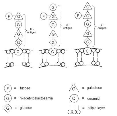
Human’s blood group is defined by the antigen properties of erythrocytes. Antigens A and B are glycoproteins, which consist 75% of carbohydrates, 15% of amino acids and 10% of phospholipids. Antigen properties depend on the nature of the sugar in glycoproteins, in other words peculiarity of antigen is defined by its carbohydrate part.
Patients with O-group (I) have antigen H, which has three carbohydrate tails (N-acetylgalactosamine, galactose, fucose).
Erythrocytes of II group have fourth tail, connected to previous - N-acetylgalactosamin.
Erythrocytes of III group have fourth tail, connected to previous - galactose.
In erythrocytes of IV group part of glycoproteins ends with galactose and part ends with N-acetylgalactosamin (fig. 3.3).
There are two sites in genome of the cell, which is responsible for blood formation – H and ABO, which are responsible for the synthesis of gene.
Figure 3.3 - Structure of glycoprotein of the erythrocyte membrane
In H-site one antigen is formed:
H-gene, which encodes enzyme fucosyl-transferase that transports fucose to galactose and controls synthesis of antigen H.
In ABO-site three antigens are formed:
Gene-O, which encodes protein that does not have fermentation activity.
A-gene, which encodes enzyme A specific transferase that transports N-acetylgalactosamine to fucose, subsequently antigen A is formed on the erythrocyte membrane.
B-gene, which encodes enzyme B specific transferase that transports galactose to fucose, subsequently antigen B is formed on the erythrocyte membrane.