A dramatic increase in the role of imaging in diagnosis of neurologic diseases occurred with the development of computed tomography (CT) in the early 1970s and of magnetic resonance imaging (MRI) in the 1980s. MRI has gradually replaced CT for many indications and has also replaced many of the invasive neuroimaging techniques, such as myelography and angiography. In general, MRI is more sensitive than CT for the evaluation of most lesions affecting the brain and spinal cord parenchyma. CT is more sensitive than MRI for visualizing osseous detail and brain hemorrhage (parenchymal or subarachnoid). Recent developments, such as helical CT, CT angiography (CTA), MR angiography (MRA), positron emission tomography, Doppler ultrasound, and interventional angiography have continued to advance diagnosis and therapy.
Computed Tomography. The CT image is a computer-generated cross-sectional representation of anatomy created by an analysis of the attenuation of x-ray beams passed through various points around a section of the body. As the x-ray source, collimated to the desired slice thickness, rotates around the patient, sensitive x-ray detectors aligned 180o from the source detect x-rays attenuated by the patient's anatomy. A computer calculates a “back projection” image from the 360o x-ray attenuation profile. Greater x-ray attenuation, as caused by bone, results in areas of high “density”, while soft tissue structures, which attenuate x-rays less, are lower in density. The density is measured in Hounsfield units (HU). The resolution of an image depends on the radiation dose, the collimation (slice thickness), the field of view, and the matrix size of the display. A typical modern CT scanner is capable of obtaining sections 1 to 2, 5, and 10 mm thick at a speed of 1 to 3 s per section; complete studies of the brain can be completed in 2 to 3 min. Intravenous contrast is often administered prior to or during a CT study to identify vascular structures and to detect defects in the blood-brain barrier (BBB) associated with pathologies such as tumors, infarcts, and infections. An intact BBB prevents contrast molecules from exiting the intravascular compartment. In the normal CNS, only vessels and those structures not having a BBB (e.g., the pituitary gland, choroid plexus, and dura) enhance. The use of contrast agents carries a risk of allergic reaction, increases the dose of radiation when both noncontrast and contrast CT scans are to be obtained, adds expense, and may mask hemorrhage; thus before contrast is administered, the indication for its use should always be considered carefully.
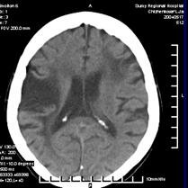
Figure 9.2. a) CT-scan. Ischemic stroke (Sumy, 2006)
Figure 9.2. b) CT-scan. Brain hemorrhage (Sumy, 2006)
Brain Magnetic Resonance Imaging (MRI). The phenomenon of magnetic resonance is a complex interaction between protons in biologic tissues, a static and alternating magnetic field (the magnet), and energy in the form of radiofrequency waves of a specific frequency (Rf), introduced by coils placed next to the body part of interest. The energy state of the hydrogen protons is transiently excited. The subsequent return to equilibrium (relaxation) of the protons results in a release of Rf energy (the echo) which can be measured by the same surface coils that delivered the Rf pulses. The complex Rf signal or echo is transformed by Fourier analysis into the information used to form an MR image.
MRI provides better resolution of neural structures than CT. This difference is most significant clinically for visualizing brain stem lesions and other abnormalities of the posterior fossa; CT images of this region are often marred by bony streak artifacts. Also, MRI is better for detecting demyelinating plaques, early infarction, subclinical brain edema, cerebral contusions, incipient transtentorial herniation, abnormalities of the craniocervical junction, and syringomyelia. It is especially valuable for identifying spinal abnormalities (e.g., tumor, abscess) compressing the spinal cord and requiring emergency intervention.
MRI is contraindicated in patients who have had a pacemaker, cardiac or carotid stents ferromagnetic aneurysm clips or other metallic objects that may overheat or be displaced within the body by the intense magnetic field.
Magnetic Resonance Angiography. Magnetic resonance angiography (MRA) uses MRI with or without a contrast agent to show cerebral vessels and major arteries and their branches in the head and neck. Although MRA has not replaced cerebral angiography, it is used when cerebral angiography cannot be done (the patient refuses or has increased risk).
Magnetic resonance venography uses MRI to show the major veins and dural sinuses of the cranium. MRV obviates the need for cerebral angiography in diagnosing cerebral venous thrombosis and is useful for monitoring thrombus resolution and guiding the duration of anticoagulation. Magnetic resonance spectroscopy can measure metabolites in the brain regionally to distinguish tumors from abscess or stroke.
Positron Emission Tomography. Positron emission tomography (PET) relies on the detection of positrons emitted during the decay of a radionuclide that has been injected into a patient. Images reveal differences in regional radionuclide activity among normal and pathologic brain structures. PET scanning has been used to assist in differentiating radiation necrosis from active neoplasm following therapy, in localizing temporal lobe epileptic foci, and in detecting metastatic disease and determining cardiac viability. Positron emission tomography (PET) is a technique which produces a three-dimensional image or picture of functional processes in the body. The system detects pairs of emitted indirectly by a emitting (tracer), which is introduced into the body on a biologically active molecule. More frequently are used FDG (Fluorodeoxyglucose). Its full chemical name is 2-fluoro-2-deoxy-D-glucose) Images of tracer concentration in 3-dimensional space within the body are then reconstructed by computer analysis.